EG:BACH7M4.5


transcription factor - basic leucine zipper - gap gene - regulates ecdysone production through specification of the PTTH-producing neurons
Please see the JBrowse view of Dmel\gt for information on other features
To submit a correction to a gene model please use the Contact FlyBase form
AlphaFold produces a per-residue confidence score (pLDDT) between 0 and 100. Some regions with low pLDDT may be unstructured in isolation.
Gene model reviewed during 5.51
1.8 (unknown)
1.9 (northern blot)
There is only one protein coding transcript and one polypeptide associated with this gene
448 (aa); 49 (kD predicted)
Homodimer or heterodimer.
Phosphorylated at multiple sites.
Click to get a list of regulatory features (enhancers, TFBS, etc.) and gene disruptions (point mutations, indels, etc.) within or overlapping Dmel\gt using the Feature Mapper tool.
The testis specificity index was calculated from modENCODE tissue expression data by Vedelek et al., 2018 to indicate the degree of testis enrichment compared to other tissues. Scores range from -2.52 (underrepresented) to 5.2 (very high testis bias).
Comment: maternally deposited
Comment: anlage in statu nascendi
Comment: anlage in statu nascendi
Comment: anlage in statu nascendi
Comment: anlage in statu nascendi
Comment: anlage in statu nascendi
Comment: reported as procephalic ectoderm anlage in statu nascendi
Comment: reported as procephalic ectoderm anlage in statu nascendi
Comment: reported as procephalic ectoderm anlage in statu nascendi
Comment: reported as procephalic ectoderm anlage
Comment: reported as procephalic ectoderm anlage
Comment: reported as procephalic ectoderm anlage
Comment: reported as procephalic ectoderm anlage
Comment: reported as procephalic ectoderm primordium
Comment: reported as procephalic ectoderm primordium
Comment: reported as procephalic ectoderm primordium
Comment: reported as procephalic ectoderm primordium
Comment: reported as procephalic ectoderm primordium
Comment: reported as procephalic ectoderm primordium
Comment: reported as crystal cell specific anlage
Comment: reported as procephalon primordium
Comment: reported as procephalon primordium
Comment: reported as procephalon primordium
Comment: reported as procephalon primordium
Comment: reported as procephalon primordium
Comment: reported as procephalon primordium
Comment: reported as plasmatocytes anlage
gt mRNA is distributed asymmetrically with respect to its protein product in embryos. The posterior border of the gt mRNA domain is located anteriorly relative to the protein border. The posterior gt mRNA reaches its maximum expression in early cycle 14A and then sharply declines, while protein levels remain high until gastrulation.
gt expression exhibits dynamic changes during nuclear cycle 14. It moves from two large domains to four stripe-like domains, while the seven striped pair-rule patterns are progressively being formed. However, the spatial relationships between gt boundaries and specific pair-rule stripes boundaries remain almost unchanged. Thus, in the anterior region, gt is always separated from run 2, ftz 2, and h 3, while it adjoins eve 2. In the posterior region, gt is separated from h 5, it adjoins eve 5, and it partially overlaps with run 5 and ftz 5.
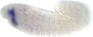
gt is expressed at the anterior lip of the ventral furrow, and in three stripes on the embryonic head, corresponding to the future ventral furrow lip, the future epipharynx and the future hypopharynx.
gt transcript is detected in three domains, one which spans between 20% and 40% egg length in the posterior, and two in the anterior, one of which reaches 10% egg length and one between 15% and 50% egg length.
gt transcripts are expressed predominantly in 2-4 hr embryos. They are expressed in two broad regions in embryonic cycle 12 extending from 60-82% egg length and 0-33% egg length. Later the posterior band narrows and a new anterior band forms from 91-97% egg length. The pattern resolves into 4 stripes, 5-6 cells wide at the cellular blastoderm stage. Cells in these stripes of expression become part of the clypeolabrum, procephalic lobe, anterior midgut invagination, and cephalic furrow. The pattern of gt expression in the posterior end of the embryo is altered in Kr, hb, and kni mutants. No change in pattern was seen in eve mutants.
gt mRNA is distributed asymmetrically with respect to its protein product in embryos. The posterior border of the gt mRNA domain is located anteriorly relative to the protein border. The posterior gt mRNA reaches its maximum expression in early cycle 14A and then sharply declines, while protein levels remain high until gastrulation.
In stage 14 embryos, gt is expressed in two neurons in the lateral portion of each brain lobe the may be either the PG neurons or precursors to the PG neurons. This expression fades by late embryogenesis. No evidence for gt expression in larval or embryonic prothoracic glands or in larval PG neurons was observed.
gt protein is first detected at the end of nuclear cycle 12 in nuclei in two distinct domains of the embryo. By cellular blastoderm, the pattern has evolved into expression in one posterior and three anterior stripes. After cellular blastoderm, gt continues to be expressed in the head region in parts of the maxillary and mandibular segments as well as in the labrum. At stage 9, transient expression is seen in the pole cells. After stage 9, all gt expression is restricted to regions of the embryo anterior to the posterior boundary of the maxillary segment. Expression persist in certain head structures including the ring gland until the end of embryonic development.
JBrowse - Visual display of RNA-Seq signals
View Dmel\gt in JBrowse

Please Note FlyBase no longer curates genomic clone accessions so this list may not be complete
Please Note This section lists cDNAs and ESTs that fall within the genomic extent of the gene model, which may include cDNAs and ESTs of genes within introns, or of overlapping genes. Please see JBrowse for alignment of the cDNAs and ESTs to the gene model.
For each fully sequenced cDNA the DGRC maintains various forms of the cDNA (e.g tagged or untagged) in several different host vectors for subsequent cloning and expression in Drosophila and Drosophila cell lines.
polyclonal
DNA-protein interactions: genome-wide binding profile assayed for gt protein in 2-3 hr embryos; see BDTNP1_TFBS_gt collection report.
S2 cells treated with dsRNA generated against this gene show reduced phagocytosis of Candida albicans compared to untreated cells.
Mutations in gt cause defects in the labial segment (the most posterior cephalic segment), and in the labral segment (the most anterior cephalic segment, adjacent to the unsegmented anterior tip).
Mutant embryos lack the corpus cardiacum.
In a sample of 79 genes with multiple introns, 33 showed significant heterogeneity in G+C content among introns of the same gene and significant positive correspondence between the intron and the third codon position G+C content within genes. These results are consistent with selection adding against preferred codons at the start of genes.
Gene product is known to regulate Kr CD (cis acting control element) expression.
Substitution of 5 amino acids in the vertebrate VBP bZIP factor that differ between the Drosophila gt bZIP factor and vertebrate PAR bZIP factors indicates that the fork region, which bridges the basic and leucine zipper domains, contributes to half-site specificity.
Muscle phenotype of mutants studied using polarised light microscopy and antibody staining to detect Mhc-lacZ reporter gene expression in muscles.
Expression of prd depends on activation by gap gene hb, Kr, kni and gt products. Primary pair rule gene products act primarily in subsequent modulation rather than activation of prd stripes. Factors activating prd expression in the pair rule mode interact with those activating it along the dorso-ventral axis.
Examination of conserved tracts in the D.virilis and D.melanogaster h gene promoters identified potential ftz-f1, Kr and gt binding sites. Kr and gt products establish the anterior and posterior borders of h stripe 5, respectively, through spatial repression.
The gene products of bcd, hb, Kr and gt all bind within the 480bp region that is necessary and sufficient for the expression of eve stripe 2. gt is involved in refining the anterior border of the stripe.
Anterior gt expression is bcd-dependent and not repressed by maternal hb. Posterior expression is independent of bcd and repressed by hb. These results strongly suggest that anterior and posterior domains of gt expression are controlled by two separate regulatory elements that act independently and respond to different regulatory proteins. gt has a strong negative effect on kni expression.
Regulatory interactions of gap genes modulate gt expression but are not required for its initiation in the embryo.
Mutations in zygotic cardinal gene gt interact with RpII140wimp.
Zygotically active locus involved in the terminal developmental program in the embryo.
gt mutants exhibit deletions of the head and abdomen.
The gt transcription unit has been identified: the temporal and spatial pattern of expression investigated. Mutants in Kr, kni and hb affect the posterior gt expression domain but mutants in eve do not affect gt expression.
Mutant embryos exhibit normal Dfd expression.
Germ line clonal analysis indicates that gt has no maternal effect.
gt mutants display defects in head and in the fifth through seventh abdominal segment.
Used by Bridges (1935) in the construction of salivary chromosome maps. Preliminary evidence of interallelic complementation with respect to phenotype and viability presented by Duttagupta, Das and Dutta (1984).
Larval development 4 days longer than normal resulting in giant larvae, pupae, and imagos. Adult weight 1.7 times normal; increased size caused by increase in cell size and not cell number (Simpson and Morata, 1980). Pupariation delayed owing to delayed increase in ecdysteroid titers; level reached at pupariation lower than normal; pupal interval of normal length (Schwartz, Imberski and Kelly, 1984). Not all genetically giant flies show the giant character, the rest have normal size; distribution sharply bimodal. Percentage giant greatest in well-fed cultures, also raised by modifying action of bb11. Penetrance of viable alleles enhanced in heterozygotes with lethal alleles and deficiencies; viability decreased (Kaufman, 1972). Abnormalities in DNA metabolism found in homo- or heteroallelic third instar gt females (Narachi and Boyd, 1985). Salivary gland chromosomes of double thickness in some cells (Bridges, 1935). Feulgen staining shows extra round of DNA synthesis; polytene chromosome can be analyzed in gt/Df larvae (Kaufman, 1972). Embryos carrying lethal giant mutations have defects in the anterior and the posterior domains (Petschek, Perrimon and Mahowald, 1987; Mohler, Eldon and Pirrotta, 1989). Posterior compartment of the labial segment deleted from blastoderm; cell death at germ-band elongation deletes anterior compartments of abdominal segments 5-7. Posterior-compartment structures of A5-7 in the peripheral nervous system fuse in mature embryos (Petschek and Mahowald, 1980). Hemizygotes for lethal alleles, gt13z and gtX11, fail to hatch (Kaufman, 1973); denticle belts of the fifth through the seventh abdominal segments partially or completely absent; internally corresponding neuromeres absent; eight abdominal neuromeres disconnected from remainder. Head does not complete involution, shows characteristic 'buttonhead' phenotype with ventral skeleton extruded from the anterior end of the larva (Mohler, Eldon and Pirrotta, 1989), pharynx and pharyngeal chitinized sclerites shorter and brain lobes somewhat smaller than normal; extensive cell death in epidermal and neural components of embryo (Honisch and Campos-Ortega, 1982). Effect cell autonomous in mosaic embryos. (Gergen and Wieschaus, 1986). Germline clones viable (Garcia-Bellido and Robbins, 1983). gt stocks (homo- or heteroallelic) show an increase in the frequency of spontaneous mutations, including deletions of y and w loci (Green, 1982); these stocks also synthesize DNA of a reduced molecular weight and show many single-strand and double-strand breaks, suggesting abnormalities in DNA metabolism (Narachi and Boyd, 1985).
Wieschaus et al. (1984) report eight embryonic lethal alleles, of which four are weak; these exhibit defects in head and in fifth through seventh abdominal segments.
Source for identity of: gt CG7952
Named "giant" because mutant embryos are slightly larger.


