msh, muscle segment homeobox, muscle specific homeobox, msh1, Dr/msh


transcription factor - homeodomain - confers cell fate on the dorsal (lateral-most) column of neuroblasts during ventral cord development - patterning of the wing disc, specification of myoblasts, proper development of muscle, neuronal and glial cells, male genital disc, and regulation of glucose metabolism
Please see the JBrowse view of Dmel\Dr for information on other features
To submit a correction to a gene model please use the Contact FlyBase form
AlphaFold produces a per-residue confidence score (pLDDT) between 0 and 100. Some regions with low pLDDT may be unstructured in isolation.
Gene model reviewed during 5.50
2.7 (northern blot)
There is only one protein coding transcript and one polypeptide associated with this gene
515 (aa)
437 (aa); 48 (kD predicted)
Click to get a list of regulatory features (enhancers, TFBS, etc.) and gene disruptions (point mutations, indels, etc.) within or overlapping Dmel\Dr using the Feature Mapper tool.
The testis specificity index was calculated from modENCODE tissue expression data by Vedelek et al., 2018 to indicate the degree of testis enrichment compared to other tissues. Scores range from -2.52 (underrepresented) to 5.2 (very high testis bias).
Comment: anlage in statu nascendi
Comment: anlage in statu nascendi
Comment: reported as procephalic ectoderm anlage in statu nascendi
Comment: reported as procephalic ectoderm anlage in statu nascendi
Comment: reported as procephalic ectoderm anlage in statu nascendi
Comment: reported as procephalic ectoderm anlage
Comment: reported as procephalic ectoderm anlage
Comment: reported as procephalic ectoderm anlage
Comment: reported as procephalic ectoderm anlage
Comment: reported as ventral nerve cord anlage
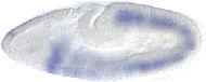
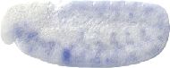
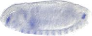
Neurectodermal expression of msh begins in embryonic stage 5 as discontinuous patches in several segments that later extend and merge to form stripes the length of the embryo. The msh-expression domain corresponds roughly to the dorsal half of the neurectoderm. From this region, 4 S1 neuroblasts of the lateral column delaminate and strong msh expression is seen in only one of these. This expression is transient and disappears by stage 9. At stage 10, expression is seen in many dorsal S3-S5 neuroblasts and in their putative neurectodermal proneural clusters. Expression is also seen in some of their immediate progeny. All are from the dorsal half of the neurogenic ectoderm but not all neuroblasts in this region express msh.
msh transcripts are detected at all stages tested except 0-2hr embryos on northern blots. The highest levels of expression are seen in embryos. A detailed description of the spatial pattern of expression in the embryo is given. In summary, expression is first observed at the cellular blastoderm stage in the lateral ectoderm and evolves into a pattern of eight stripes. The lateral expression prefigures the appearance of lateral proneural clusters of the CNS. During the first wave of neuroblast segregation, expression is restricted to delaminating neuroblasts (1 in 4 lateral neuroblasts per hemisegment express msh) and is excluded from ectodermal cells. Subsequent waves of neuroblast segregation are also preceded by msh expression in cell clusters in the the lateral neurectoderm. msh is also expressed in sensory organ precursors of the PNS. Three additional sites of msh in the embryonic ectoderm include the precursors to the stomatogastric neurons, the region that will give rise to neurons of the brain and optic lobes, and the salivary gland placodes. msh expression is seen in dorsal mesodermal cells that will give rise to dorsal body wall muscles and ventral-lateral mesodermal cells that will give rise to fat body. In wing discs, msh is expressed in a highly restricted pattern. Highest expression occurs in three patches of cells in the prospective dorsal hinge region, two of which include proneural clusters. Areas of weaker expression include a row of cells that will develop into sensilla of the anterior wing margin.
msh transcripts are detected at all stages tested except third instar larvae on northern blots. A peak of expression occurs in 8-12hr embryos. By in situ hybridization, transcripts are first detected at stage 5 of embryogenesis in a bilateral series of segmentally repeated bands of dorsal lateral ectodermal cells. At around stage 8, this is replaced with a transient ventral lateral band of msh expression extending along most of the germ band. In stage 10, expression is seen in segmentally repeated clusters of neuroblasts in the thoracic and abdominal segments. By stage 12, in addition to strong expression in segregated neuroblasts, expression is seen in clusters of mesodermal cells. By stage 13, expression is primarily in the CNS and in the developing somatic musculature (corresponding mostly to the developing dorsal and lateral somatic body wall muscles). Expression is also seen in the foregut, hindgut, and fat body.
Comment: lateral posterior
Dr protein is expressed in the male genital discs of third instar larvae. Expression is localized to a medial domain on the ventral surface of the A9 primordium encompassing the bnl-expressing cells but extending laterally and posteriorly; and (2) a subset of the btl-expressing cells that are adjacent to those expressing bnl.
At embryonic stage 15, Dr expression is seen in specific cell clusters in each brain neuromere. Protcerebral Dr expression begins at stage 12 and continues through embryogenesis. A domain of expression is observed in the deutocerebrum consisting of about 30 cells, which are mainly located near and within the protocerbral-deuterocerebral boundary. Another expression domain is located in the tritocerebrum distributed along the entire neuromere. Dr expression is also observed in cells closely associated with the preoral commissure and cervical as well as longitudinal axon tracts. Expression is also observed in specific cell clusters at the mediolateral margin of each individual neuromere in the posterior brain and ventral nerve cord. Expression is also observed in cell bodies associated with the paired longitudinal connectives, resembling longitudinal glial cells.
Expression in procephalic neuroblasts stage 9-11: tritocerebrum - d4-8; deuterocerebrum - d2-13
JBrowse - Visual display of RNA-Seq signals
View Dmel\Dr in JBrowse

3-99
3-96.4
3-99.2
Please Note FlyBase no longer curates genomic clone accessions so this list may not be complete
Please Note This section lists cDNAs and ESTs that fall within the genomic extent of the gene model, which may include cDNAs and ESTs of genes within introns, or of overlapping genes. Please see JBrowse for alignment of the cDNAs and ESTs to the gene model.
For each fully sequenced cDNA the DGRC maintains various forms of the cDNA (e.g tagged or untagged) in several different host vectors for subsequent cloning and expression in Drosophila and Drosophila cell lines.
"Dr" mutants are always double hits in two genes, one of which is "stg".
Dr is expressed in the dorsal and lateral domains of muscle progenitors and is required for the specification of the progenitor cells. Ectopic expression of Dr in the entire mesoderm inhibits the proper development of the normally Dr-negative muscle progenitors in the dorsolateral domain. Results suggest Dr plays a role in regional specification of muscle progenitors/founders.
Ectopic expression of Dr in the entire neuroectoderm severely disrupts the development of the midline and ventral neuroblasts.
Dr is required for the correct development of dorsal neuroblasts in the embryo.
Expression of Dr in the neuroectoderm prefigures the formation of proneural clusters. Dr expression also prefigures invagination centres of the stomatogastric nervous system. Dr is regulated by the dorsoventral patterning genes Egfr and dpp in early embryos. Ectodermal and mesodermal Dr expression depend on wg and hh.
Normal expression and the effects of ectopic expression of Dr gene suggest a role in differentiation and patterning of embryonic muscles.
The Dr homeobox sequence has been used as a probe to isolate a homologous mouse gene, called Hox-7.
The DNA sequences of the homeobox region of 11 Drosophila genes, including Dr, have been compared.
Heterozygotes have extremely reduced numbers of facets, 1-10 in the case of Dr1 and DrA; about 30 facets coalesce to give shiny, dark red appearance in DrMio; five to seven rhabdomeres per ommatidium section; some rhabdomeres fuse. Developing eye disc reduced in size; morphogenetic furrow uneven; few clusters of presumptive photoreceptor cells; arrangement disturbed; microvillar caps blurred and diffuse (Renfranz and Benzer, 1989).
Source for merge of: ltt msh
Source for merge of: Dr msh
"Dlw" mutations may be regulatory mutants of the Dr gene; "Dr" gain-of-function mutants contain lesions in the same region as "Dlw" mutants (several kb upstream of the transcription start site), DrΔ68 is lethal in combination with Dlw1, Dlw3 or Dlw4 and the level of Dr mRNA is reduced throughout the wing pouch in Dlw1 mutants (which may account for the loss of function characteristics seen in homozygous Dlw1 mutant clones).
Source for identity of: msh CG1897
'Lottchen' is the name of twins in the book 'Das doppelte Lottchen' by Erich Kaestner.


